HPHT vs CVD Diamonds: Principles, Differences, and Identification Guide”
Table of Contents
With the continuous advancement of gemological technology, synthetic diamonds (commonly referred to as lab-grown diamonds) have moved from laboratory research into the commercial market. Today, the two primary methods for growing diamonds in a laboratory setting are High Pressure High Temperature (HPHT) and Chemical Vapor Deposition (CVD).
While both types share identical physical, chemical, and optical properties with natural diamonds, their growth environments leave distinct microscopic characteristics and defect structures. Understanding these characteristics is essential for gemological testing and accurate identification.
1. Growth Principles of Synthetic Diamonds
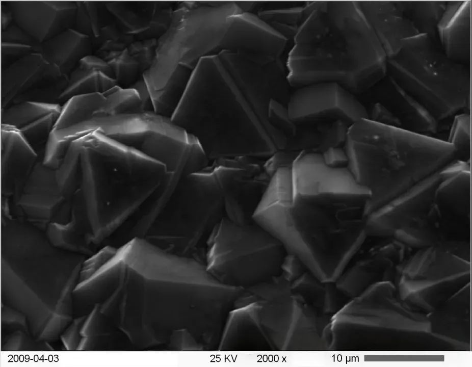
High Pressure High Temperature (HPHT) Growth Principle
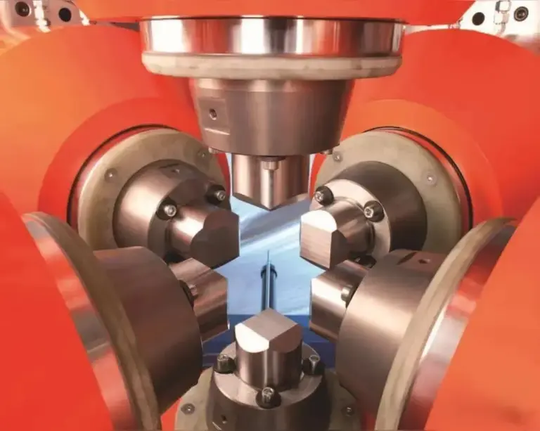
The HPHT method simulates the extreme temperature and pressure conditions found deep within the Earth’s mantle where natural diamonds form.
- Growth Environment: Temperatures typically range from 1,300 degrees Celsius to 1,600 degrees Celsius, with pressures reaching 5 to 6 GPa
- Equipment: Specialized high-pressure apparatuses are used, such as cubic presses, belt presses, or split-sphere (BARS) systems.
- Mechanism: A carbon source (such as graphite) is placed in the high-temperature zone, while a diamond seed crystal is placed in the lower-temperature zone. Metal catalysts—commonly iron (Fe), nickel (Ni), or cobalt (Co)—are used to lower the required growth temperature and pressure. The carbon dissolves into the molten metal flux and migrates toward the seed crystal, depositing layer by layer to form a synthetic diamond crystal.
.
Chemical Vapor Deposition (CVD) Growth Principle
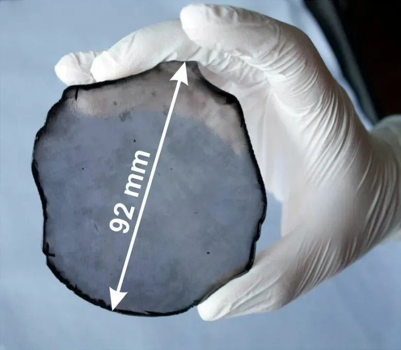
The CVD method synthesizes diamonds under low pressure and high temperature by triggering chemical gas-phase reactions.
Growth Environment: Temperatures range between 700°C and 1200°C, under sub-atmospheric pressures (typically below 100 Torr).
Mechanism: Methane (CH₄) and hydrogen (H₂) serve as the primary reaction gases. An energy source—most commonly Microwave Plasma (MPCVD)—is used to dissociate the gas molecules into reactive radicals, including atomic carbon and hydrogen ions. Atomic hydrogen selectively etches away non-diamond carbon (such as graphite), allowing active carbon species to deposit epitaxially onto a single-crystal diamond substrate, growing the crystal layer by layer.
2. Structural Characteristics and Internal Defects
Due to the fundamental differences in their growth mechanisms, HPHT and CVD lab-grown diamonds exhibit distinct structural features, growth patterns, and internal defects.
Morphological and Growth Sector Differences
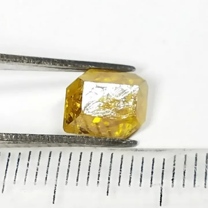
- HHPHT Diamonds: Exhibit a multi-directional cuboctahedral growth morphology, displaying distinct growth sectors such as octahedral {111}, cubic {100}, and dodecahedral {110} faces.
- CVD Diamonds: Typically follow a single-direction tabular growth pattern, growing predominantly along the {100} crystallographic direction.
Inclusions and Internal Features
- HPHT Inclusions: Frequently contain metallic flux inclusions composed of Fe, Ni, or Co. These inclusions often display a metallic luster, tabular or pin-like shapes, and can render the diamond magnetic.
- CVD Inclusions: Mainly contain non-diamond carbon particles (such as graphite or amorphous carbon) and dark pinpoint inclusions. They do not contain metallic flux inclusions.
Impurities and Atomic Defect Structuresv
- Nitrogen Impurities:
- HPHT: Nitrogen is predominantly present in the isolated single-atom state (C-center, Type Ib diamond). Nitrogen-vacancy centers (NV⁻ and NV⁰) may also be introduced during or after growth.
- CVD: Nitrogen content is usually very low (often Type IIa diamond). However, deliberate nitrogen doping can create NV centers, imparting a brown hue or specific optical features.
- Silicon Impurities: CVD diamonds frequently exhibit silicon-vacancy centers ([Si-V]⁻) caused by silicon contamination from the quartz vacuum chambers used during manufacturing. This defect is rare in HPHT diamonds.
3. How to Identify HPHT and CVD Lab-Grown Diamonds
Accurately distinguishing between natural, HPHT, and CVD diamonds requires a combination of standard gemological testing and advanced laboratory spectroscopic analysis.
Standard Gemological Inspection
Fluorescence and PhosStandard Gemological Inspection
- Microscopic Observation:
- Examine for metallic flux inclusions (HPHT) or dark graphite pinpoint clusters (CVD).
- Observe color distribution and sectorial zoning; HPHT diamonds often exhibit geometric color zoning corresponding to specific growth sectors.
- phorescence:
- HPHT Diamonds: Frequently exhibit strong short-wave ultraviolet (SWUV) fluorescence and long-lasting phosphorescence, often displaying green, yellow-green, or blue hues.
- CVD Diamonds: Typically show weak to moderate fluorescence under long-wave UV (LWUV), but may display distinctive orange-red or pinkish-orange fluorescence under SWUV due to high concentrations of $\text{NV}$ centers.
Strain Patterns (Anomalous Birefringence):
- CVD Diamonds: Display characteristic “columnar” or “banded” strain patterns under cross-polarized light due to stress accumulated during high-rate layer-by-layer growth.
- HPHT Diamonds: Generally exhibit very low strain levels or weak, localized strain patterns.
Advanced Spectroscopic Analysis
- Photoluminescence (PL) Spectroscopy:
- $[\text{Si-V}]^-$ Defect Peak ($737\text{ nm}$): A crucial indicator for CVD diamonds.
- Nickel/Cobalt-Related Peaks ($883\text{ nm} / 884\text{ nm}$): Characteristic optical features seen in HPHT diamonds grown with Ni-based flux.
- NV Centers ($575\text{ nm}$ and $637\text{ nm}$): Used to evaluate nitrogen content and post-growth annealing treatments in both HPHT and CVD diamonds.
- Fourier-Transform Infrared (FTIR) Spectroscopy:
- Used to classify diamond type (Type Ia, Ib, IIa, IIb). Most CVD and many high-purity HPHT diamonds are classified as Type IIa (nitrogen-free or virtually nitrogen-free).
- DiamondView™ Deep-UV Fluorescence Imaging:
- HPHT Diamonds: Reveal distinct geometric, cross-shaped, or sectorial fluorescence growth structures.
- CVD Diamonds: Display layered, parallel growth lines reflecting their step-by-step planar deposition process.
4. Summary
While HPHT and CVD technologies continue to evolve—producing larger, higher-clarity, and colorless lab-grown diamonds—their fundamental physical growth mechanisms remain inherently different.
By analyzing microscopic inclusions, UV fluorescence/phosphorescence behaviors, birefringence strain patterns, and specific spectroscopic features (such as [Si-V]⁻ and [Ni-V] defects), gemological laboratories can reliably identify and differentiate HPHT and CVD lab-grown diamonds from their natural counterparts.